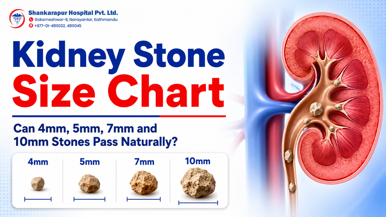
Kidney stone size strongly influences whether a stone may pass naturally, but size alone cannot predict the outcome. A 4mm stone in the lower ureter may pass without a procedure, while a 5mm stone causing infection or obstruction may require urgent treatment. A 7mm stone can sometimes pass, although the chance becomes less predictable as size increases. A 10mm stone is less likely to pass easily and usually needs assessment by a urologist.
Stone location, pain, infection, urine blockage, kidney function and overall health are also important. This kidney stone size chart explains what different measurements may mean and when observation, medicines, laser treatment, shock wave treatment or surgery may be considered.
Important: The chart provides general educational guidance. A treatment decision should be based on imaging, symptoms, urine and blood tests, kidney function and a urologist’s evaluation.
Kidney Stone Size Chart at a Glance
The following chart gives a general interpretation of stone sizes. Natural-passage estimates mainly apply to stones that have already moved into the ureter; the tube connecting the kidney to the bladder.
| Kidney stone size | General interpretation | Possibility of natural passage | Common next step |
|---|---|---|---|
| Under 4mm | Small stone | Often more likely to pass if it is in the ureter and no complication is present | Monitoring, symptom control and follow-up |
| Around 5mm | Small to moderate stone | Passage may still occur, but location becomes important | Urologist-guided observation or treatment planning |
| 6–7mm | Moderate stone | Passage is possible but less predictable | Closer monitoring and urology assessment |
| 8–10mm | Moderate to larger stone | Less likely to pass easily | Active treatment may be discussed |
| 10–20mm | Larger stone | Natural passage is generally less likely | ESWL, URS, RIRS or another procedure may be considered |
| Over 20mm | Large stone burden | Unlikely to pass naturally | PCNL or another planned procedure is commonly considered |
The European Association of Urology reports that available passage data are limited, but approximately 75% of ureteric stones smaller than 5mm and 62% of stones measuring 5mm or more passed spontaneously in the studies it reviewed. The likelihood decreased as stone size increased and also differed according to the stone’s location. These percentages should not be treated as a personal prediction.
Is a Kidney Stone the Same as a Ureteric Stone?
A kidney stone forms inside the kidney. A ureteric stone is a kidney stone that has moved into the ureter.
This difference matters because most natural-passage data relate to ureteric stones. A stone still sitting inside the kidney may remain there without symptoms, grow over time or move into the ureter later. It does not begin its full passage toward the bladder until it enters the ureter.
That is why two people with the same “7mm stone” on a report may need different plans:
-
One may have a non-obstructing stone inside the kidney.
-
Another may have a stone stuck in the ureter.
-
One may have no pain or infection.
-
Another may have kidney swelling, severe pain or fever.
Stone size should always be interpreted together with its exact location.
Can a 4mm Kidney Stone Pass Naturally?
A 4mm ureteric stone often has a reasonable possibility of passing naturally, particularly when it is closer to the bladder. However, natural passage is not guaranteed.
Observation may be considered when pain can be controlled, urine is flowing normally, there is no significant infection and kidney function is stable. Follow-up matters because symptoms can improve even when a stone has not completely passed.
A 4mm stone may still need active treatment when it causes:
-
Persistent or severe pain
-
Fever or urinary infection
-
Ongoing urine blockage
-
Kidney swelling
-
Reduced kidney function
-
Repeated emergency visits
-
Failure to move during follow-up
Do not assume that every 4mm stone is harmless simply because it is small.
Can a 5mm Kidney Stone Pass Naturally?
A 5mm stone may pass naturally, but the outcome depends heavily on its location and the patient’s condition. A stone near the bladder generally has a better chance of passing than a similarly sized stone in the upper ureter.
The measurement itself can also vary slightly between ultrasound, X-ray and CT reports. A stone reported as 5mm should therefore not be treated as an exact guarantee of natural passage.
A urologist may recommend monitoring for a selected 5mm ureteric stone when:
-
Symptoms are manageable
-
There is no fever or significant infection
-
Urine flow is not dangerously obstructed
-
Kidney function is stable
-
Follow-up imaging is possible
Prescription treatment intended to support stone passage may be considered for selected patients, but it should only be used after professional assessment.
Can a 7mm Kidney Stone Pass Without Surgery?
A 7mm ureteric stone can sometimes pass, but it is less predictable than a stone smaller than 5mm. Location, urinary anatomy and the presence of obstruction can substantially change the likelihood.
A 7mm stone in the lower ureter may have a better chance of passing than one in the upper ureter. However, waiting may not be appropriate when the stone causes persistent pain, infection, urine blockage or deterioration in kidney function.
A urologist may evaluate:
-
Whether the stone is moving
-
How long symptoms have continued
-
Whether the kidney is swollen
-
Whether infection is present
-
Whether pain medicine is controlling symptoms
-
Whether the patient has one functioning kidney
-
Whether the patient can return for follow-up
Laser ureteroscopy, RIRS, shock wave lithotripsy or another treatment may be considered when observation is unlikely to succeed or is no longer safe.
Does a 10mm Kidney Stone Need Surgery?
A 10mm kidney stone does not automatically require surgery, but it is less likely to pass naturally than a smaller ureteric stone. Urologist evaluation is important because treatment depends on whether the stone is in the kidney or ureter, whether it is causing symptoms and whether urine flow is obstructed.
For a 10mm stone inside the kidney, factors such as the stone’s position, density, growth and symptoms influence whether it is monitored or treated. For a 10mm stone lodged in the ureter, active removal may be discussed more strongly, especially when it is not progressing.
Possible treatments include:
-
Shock wave lithotripsy, or ESWL
-
Ureteroscopy with laser lithotripsy
-
Retrograde intrarenal surgery, or RIRS
-
Percutaneous nephrolithotomy, or PCNL, for selected larger or complex stones
The safest option cannot be selected from size alone.
Why Does Stone Location Matter So Much?
A stone generally has a better chance of passing as it moves closer to the bladder.
The European Association of Urology reports spontaneous-passage ranges of approximately:
| Stone location | Reported spontaneous-passage range |
|---|---|
| Upper ureter | 49–52% |
| Middle ureter | 58–70% |
| Distal or lower ureter | 68–83% |
These figures combine stones of different sizes and patient situations. They demonstrate why “How many millimetres is the stone?” is only one part of the decision.
Upper ureter
An upper ureteric stone is located closer to the kidney. It has a longer distance to travel and may be less likely to pass than a similar stone located lower down.
Middle ureter
A stone in the middle portion of the ureter may continue to progress, but follow-up is required to confirm movement and detect obstruction.
Distal or lower ureter
A distal stone is closer to the bladder. Smaller distal stones generally have a better possibility of natural passage, provided no complication is present.
Stone inside the kidney
A non-obstructing kidney stone may not cause immediate pain. Depending on its size, growth, position and the patient’s history, a urologist may recommend observation or planned treatment.
What Determines Treatment Besides Stone Size?
A safe treatment plan considers the whole clinical picture.
| Assessment factor | Why it matters |
|---|---|
| Exact stone location | Kidney and ureter stones behave differently |
| Number of stones | Multiple stones may make treatment more complex |
| Pain severity | Persistent uncontrolled pain may require intervention |
| Infection | Infection with obstruction can become an emergency |
| Urinary obstruction | Blockage can affect kidney function |
| Kidney swelling | Hydronephrosis may suggest impaired urine drainage |
| Kidney function | Reduced function may increase treatment urgency |
| Stone density | Harder stones may respond differently to shock waves |
| Stone growth | A growing stone may need active management |
| Previous treatment | Failed observation or ESWL can change the next step |
| Urinary anatomy | Anatomy affects passage and procedure selection |
| Overall health | Anaesthesia and procedural risks differ between patients |
The European Association of Urology lists a low likelihood of spontaneous passage, persistent pain, persistent obstruction and renal insufficiency among the reasons for removing a ureteric stone.
How Is Kidney Stone Size Confirmed?
Kidney-stone evaluation may involve imaging, urine tests, blood tests and clinical examination.
Ultrasound
Ultrasound can identify kidney stones, urinary-system swelling and selected stones near the upper or lower ends of the ureter. It does not use ionising radiation.
CT scan
A non-contrast CT scan can provide more detailed information about stone size, location and density. The European Association of Urology recommends non-contrast CT to confirm stone diagnosis in patients with acute flank pain after an initial ultrasound assessment when clinically appropriate.
X-ray
Some stones are visible on X-ray, while others are not. An X-ray may sometimes be used for follow-up or treatment planning.
Urine and blood tests
Tests may help assess infection, kidney function and factors associated with stone formation.
Shankarapur Hospital publicly lists CT scan, digital X-ray and ultrasound among its Diagnostic and Imaging Services.
When Can a Kidney Stone Be Monitored?
Observation may be appropriate for selected small stones when the patient is clinically stable and can return for follow-up.
Monitoring may involve:
-
Prescribed pain control
-
Adequate fluid intake when medically appropriate
-
Follow-up imaging
-
Urine straining to collect a passed stone
-
Monitoring for fever or worsening symptoms
-
Urologist-prescribed medicine in selected cases
Observation should not mean ignoring the stone. A follow-up plan is needed to confirm whether the stone has passed, remained stable or caused obstruction.
The National Institute of Diabetes and Digestive and Kidney Diseases explains that small stones may pass through the urinary tract without a procedure, while larger stones, severe pain or urinary blockage may require urgent treatment.
What Size Kidney Stone Requires Treatment?
There is no single size at which every kidney stone must be removed.
Active treatment is considered more strongly when:
-
The stone is unlikely to pass
-
Pain remains severe despite treatment
-
The stone blocks urine
-
Infection is present
-
Kidney function is affected
-
The stone continues to grow
-
Symptoms repeatedly return
-
Observation has failed
-
The patient has a single functioning kidney
-
The stone’s position makes natural passage difficult
For kidney stones larger than 20mm, the European Association of Urology recommends PCNL as the primary treatment in many cases because other methods may require multiple procedures. For stones between 10mm and 20mm, treatment selection may include RIRS, SWL or PCNL depending on location, anatomy, density, symptoms and procedural considerations.
Kidney Stone Treatment Options According to Size and Location
Observation and medical management
This may be suitable for selected small stones that are likely to pass and are not causing infection, uncontrolled pain or dangerous obstruction.
Shock wave lithotripsy
Extracorporeal shock wave lithotripsy uses energy from outside the body to break a stone into smaller fragments. Its suitability depends on stone size, location, density and urinary anatomy.
Ureteroscopy with laser lithotripsy
A thin scope is passed through the natural urinary tract to locate the stone. Laser energy may then break the stone into small pieces.
This treatment is commonly considered for ureteric stones and selected kidney stones. More detail is available in Shankarapur Hospital’s guide to laser surgery for kidney stones.
Retrograde intrarenal surgery
RIRS uses a flexible scope passed through the urinary passage to reach selected stones inside the kidney. The stone may be fragmented with laser energy.
Percutaneous nephrolithotomy
PCNL removes a stone through a small access tract created through the back. It is commonly used for large or complex kidney stones.
Shankarapur Hospital’s Urology Department lists kidney-stone management using PCNL, URS and RIRS, along with endoscopic and laser procedures.
| Treatment | Common role | Important limitation |
|---|---|---|
| Observation | Selected small, uncomplicated stones | Requires reliable follow-up |
| ESWL | Selected kidney or upper ureter stones | May require repeat treatment |
| URS with laser | Ureteric and selected kidney stones | Anaesthesia and endoscopic access are required |
| RIRS | Selected stones inside the kidney | Some stones may require staged treatment |
| PCNL | Large or complex kidney stones | More invasive than URS or RIRS |
The choice is not simply “small stone versus large stone.” A urologist must connect stone size with location, symptoms, infection, kidney function and the advantages and limitations of each procedure.
Hypothetical Example: Two Different 7mm Stones
Consider two hypothetical patients whose reports both show a 7mm stone.
Patient A has a 7mm stone inside the kidney, no pain, no infection and no urinary obstruction. The urologist may discuss observation with scheduled follow-up or planned treatment depending on the stone’s position, growth and patient preference.
Patient B has a 7mm stone lodged in the ureter, repeated severe pain, kidney swelling and fever. This situation requires urgent hospital assessment because an obstructed urinary system with infection can become serious.
The measurement is the same, but the clinical risk is completely different.
When Is a Kidney Stone an Emergency?
Seek urgent medical evaluation when kidney-stone symptoms are accompanied by:
-
Fever or chills
-
Severe pain that does not settle
-
Inability to pass urine
-
Very low urine output
-
Repeated vomiting or dehydration
-
Confusion or extreme weakness
-
Pain with a known single functioning kidney
-
Pregnancy with severe urinary symptoms
-
Worsening symptoms after a stone diagnosis
Kidney stones may cause sharp back, side, lower abdominal or groin pain, blood in urine, painful urination, urinary urgency, nausea, vomiting, fever or chills.
A blocked urinary system with infection should not be managed with home remedies or delayed while waiting for the stone to pass.
What Affects Kidney Stone Treatment Cost in Nepal?
The cost of kidney-stone treatment in Nepal varies because not every patient needs the same tests or procedure.
Cost may depend on:
-
Consultation and diagnostic tests
-
Ultrasound, X-ray or CT scan
-
Stone size and location
-
Medicine versus a procedure
-
ESWL, URS, RIRS or PCNL
-
Anaesthesia
-
Hospital admission
-
DJ stent placement and removal
-
Medicines and follow-up
-
Infection or emergency treatment
-
Need for more than one procedure
An exact estimate should be provided after the urologist reviews the patient and imaging reports. Shankarapur Hospital’s broader guide to kidney stone treatment options in Nepal explains the main treatment and cost factors.
Contextual CTA
Have you received an ultrasound or CT report showing a 5mm, 7mm or 10mm stone? Bring the report to a urology consultation so the stone’s size, location, obstruction and treatment options can be assessed together.
Can Drinking More Water Remove Every Kidney Stone?
Drinking enough fluid can help prevent concentrated urine and may support the passage of selected small stones. However, water cannot safely remove every stone.
A larger, infected or obstructing stone may not pass simply by drinking more water. Excessive fluid intake should not be used as a substitute for medical assessment when pain is severe, vomiting is present or urine flow is reduced.
Patients with kidney disease, heart disease or fluid restrictions should follow individual medical advice rather than increasing fluid intake independently.
Can Kidney Stones Return After Passing or Removal?
Yes. Removing or passing a stone does not always correct the reason it formed.
Prevention may involve:
-
Identifying the stone type
-
Maintaining suitable fluid intake
-
Reducing excessive sodium
-
Following condition-specific dietary advice
-
Managing recurrent urinary infections
-
Reviewing relevant medicines and health conditions
-
Completing blood or urine testing when advised
Do not remove all calcium-containing foods without professional advice. Prevention depends on the type of stone and the patient’s urine, diet and medical history.
Shankarapur Hospital’s article on kidney-stone causes, symptoms and prevention provides further background.
Questions to Ask a Urologist About Your Stone
Bring your imaging report and ask:
-
Is the stone inside my kidney or ureter?
-
Is it blocking urine flow?
-
Is my kidney swollen?
-
Is there evidence of infection?
-
What is the exact stone measurement?
-
Is the stone moving?
-
Is observation safe in my situation?
-
When should follow-up imaging be done?
-
What symptoms mean I should visit emergency care?
-
Which treatments are appropriate for this stone?
-
Could I need a DJ stent?
-
What is included in the treatment estimate?
-
How can the stone be tested after removal?
-
What can help reduce recurrence?
These questions create a clearer discussion than asking only, “Is my stone big?”
Frequently Asked Questions
Can a 4mm kidney stone pass naturally?
A 4mm ureteric stone often has a reasonable possibility of passing without a procedure, especially when it is in the lower ureter. However, treatment may still be required if the stone causes infection, persistent pain, obstruction or reduced kidney function. Follow-up should confirm that it has actually passed.
Can a 5mm kidney stone pass through urine?
A 5mm stone may pass through urine, but the likelihood depends on whether it is inside the kidney or ureter and how close it is to the bladder. Symptoms, obstruction and kidney function also influence whether observation is appropriate.
Can a 7mm kidney stone pass naturally?
A 7mm ureteric stone can sometimes pass naturally, but passage is less predictable than with stones smaller than 5mm. Urologist assessment is recommended to evaluate the stone’s location, movement, obstruction, pain and infection risk.
Does a 10mm kidney stone require surgery?
A 10mm kidney stone does not always require surgery, but natural passage is less likely. Treatment may include ESWL, ureteroscopy, RIRS or another procedure depending on its location, density, symptoms, obstruction and the patient’s health.
What size kidney stone is considered large?
Stone size is interpreted in context, but stones above 10mm generally need closer treatment planning, while renal stones above 20mm are considered a large stone burden and are commonly treated with PCNL. Location and clinical complications remain important.
What size kidney stone requires laser treatment?
There is no single measurement that automatically requires laser treatment. Laser ureteroscopy or RIRS may be considered when a stone is unlikely to pass, is stuck in the ureter, causes persistent symptoms or is unsuitable for observation or shock wave treatment.
How long does a kidney stone take to pass?
Passage time varies. The European Association of Urology cites an average of about 17 days in reviewed ureteric-stone data, with a reported range of 6–29 days. Individual cases may be shorter or longer, and waiting is unsafe when complications develop.
Can a kidney stone remain without causing pain?
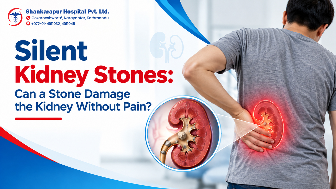
Yes. A stone inside the kidney may be discovered incidentally and cause no immediate symptoms. It may remain stable, grow or move into the ureter later. Follow-up depends on its size, location, growth and the patient’s risk factors.
Which scan shows kidney-stone size accurately?
Ultrasound can detect many kidney stones and urinary obstruction without radiation. Non-contrast CT generally provides more detailed information about stone size, location and density when clinically required. The appropriate test depends on the patient and clinical situation.
Is the stone’s location more important than its size?
Both are important. Size influences the likelihood of passage, while location influences how far the stone must travel and which treatment can reach it. A smaller upper-ureter stone and a larger lower-ureter stone may behave differently.
When should kidney-stone symptoms be treated as an emergency?
Seek urgent care for fever or chills, severe uncontrolled pain, inability to pass urine, very low urine output, repeated vomiting, worsening weakness or symptoms involving a single functioning kidney. Infection combined with urinary obstruction can become serious.
Key Takeaways
-
Stone size affects passage, but it is not the only deciding factor.
-
Stones smaller than 5mm are generally more likely to pass than larger stones.
-
A 7mm stone can pass, but the outcome is less predictable.
-
A 10mm stone usually needs urologist assessment and may require a procedure.
-
A stone’s location in the kidney or ureter changes its likely behaviour.
-
Fever, obstruction, uncontrolled pain and reduced kidney function can make treatment urgent.
-
Imaging and clinical evaluation are necessary before choosing observation, ESWL, URS, RIRS or PCNL.
-
Patients should not select treatment based only on an online chart.
A kidney-stone report should be interpreted according to more than its measurement. Shankarapur Hospital’s Urology Department provides kidney-stone evaluation and lists PCNL, URS, RIRS, endoscopic procedures and laser stone-removal services.
Bring your ultrasound, CT scan and previous test reports for an individualized urology assessment.
Book a Urology Consultation
AUTHOR
Shankarapur Hospital Editorial Team
The Shankarapur Hospital Editorial Team prepares patient-education content about health conditions, diagnostic services and hospital care. This article is intended for general education and does not replace examination, imaging or treatment advice from a qualified healthcare professional.